
Location:
- The liver is the largest gland in our body that weighs between 1 to 2.3 kg in a healthy adult.
- It is situated in the abdominal cavity in the upper right part just below the diaphragm.
- Its upper and anterior surfaces are smooth and lie in close association with the posterior surface of the diaphragm while its posterior surface is irregular in outline.
- The liver is enclosed in a thin capsule and completely covered by a thin fold of peritoneum called falciparum ligament which attaches the liver to the diaphragm to keep it in proper position.
Structure:
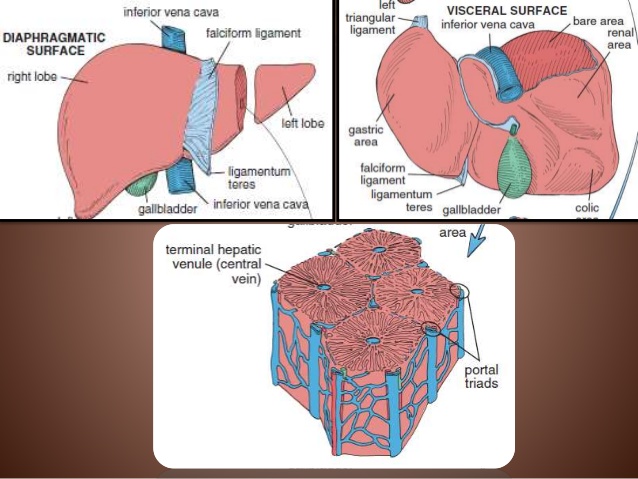
- The liver is made up of two major lobes; left and right lobes which are joined together by falciparum ligament.
- The left lobe is smaller and forms about one sixth of the liver. The right lobe is much larger and further incompletely divided by shallow fissures into three lobes;
- the larges right lobe proper,
- a small four sided caudate lobe and
- a small oblong quadrate lobe.
- To the posterior surface of liver in between the right central and quadrate lobes, is attached a pear-shaped sac-like structure called gall bladder.
- Gall bladder is dark green in color with a thin wall that stores the secretions of liver like bile.
- A large cystic duct arises from the gall bladder which receives several small hepatic ducts from different lobes of the liver carrying bile.
- A large cystic duct from gall bladder and hepatic ducts from liver collectively form a large common bile duct which opens into the proximal part of the duodenum in the small intestine. The opening is guarded by a sphincter.
Histology of liver:
- Each lobe of liver consists of a large number of hepatic lobules that are separated from each other by a thin layer of connective tissue called septa or Glisson’s capsule.
- In the liver of rabbit and human beings, septa demarcating the lobules are incomplete and not clearly marked off. The lobules are just visible to the naked eyes and are hexagonal in outline.
- In the center of each lobule lies a branch of hepatic vein, the intralobular vein or central vein.
- The cells forming lobules are cuboidal or polyhedral in shape and are called hepatic cells or hepatocytes. These are arranged in radial rows of 1 or 2 cell thick rods and trabeculae which extend from the central vein to the periphery of the lobule.
- Each liver cell possesses a fine granular cytoplasm, large round central nucleus and deposits of glycogen and lipids.
- A labyrinth of narrow, irregular spaces called lacunae is present between the hepatic cords. These lacunae have blood capillaries called hepatic sinusoids.
- The walls of the sinusoid are discontinuous and are made up of endothelial cells.
- Some of these cells become highly phagocytic and are called cells of Kupffer of Kupffer cells which ingest dead erythrocytes and destroy harmful bacteria.
- Strands of interlobular connective tissue called portal areas or portal canals are present at the corner between the adjacent lobules.
- Each portal canal supports a branch of hepatic artery, hepatic portal vein and bile duct which are often termed interlobular.
- Portal canals are also supplied with nerves and lymphatic vessels.
- Sinusoids contain blood coming from inter lobular branches of hepatic portal vein and hepatic artery and empty into the intralobular central veins which unite to form the hepatic vein.
- An intricate meshwork of fine intercellular channels of tubules called bile capillaries surrounds the hepatic cells.
- Bile capillaries receive bile directly from the liver. These liver capillaries join to form bile ductules that in turn open into hepatic ducts. Hepatic ducts ultimately form the common bile duct.
Functions of liver:
- Liver performs a number of functions, so it is also called a master laboratory of the body. It is an important organ for metabolism in vertebrates. A summary of its important functions is as follows:
- Secretion of bile:
- Hepatocytes or liver cells secrete a dark green colored alkaline fluid called bile from the mixed arterial and venous blood in the sinusoids. Bile consists of bile salts, bile pigments, cholesterol, lecithin, water etc. Bile performs the following functions in our body;
- It makes the chyme alkaline, better suited for the action of pancreatic juice.
- It brings about the emulsification of fats.
- It helps in removing the excretory product like bile pigments, inorganic salts, toxins etc. from the body.
- It checks the bacterial growth and multiplication (has an antiseptic effect).
- It helps in the absorption of fat soluble vitamins like A, D, E and K.
- It also stimulates peristalsis.
- Hepatocytes or liver cells secrete a dark green colored alkaline fluid called bile from the mixed arterial and venous blood in the sinusoids. Bile consists of bile salts, bile pigments, cholesterol, lecithin, water etc. Bile performs the following functions in our body;
- Secretion of bile:
- Deamination:
- It decomposes the excess proteins and amino acids to ammonia from which urea is formed through a chain of reactions.
- It breaks down nucleic protein of worn out cells in the body to form uric acid which is excreted in urine.
- Glycogenesis and glycogenolysis:
- It converts excess glucose into glycogen (glycogenesis) in the presence of insulin and changes glycogen to glucose (glycogenolysis) in the presence of glucagon. Thus, it helps in maintaining a constant sugar level in the blood.
- Desaturation:
- It converts stored fat to a form in which it can be used by the tissues to provide energy.
- Storage:
- It serves as a storage organ for:
- Vitamin B12
- Fat soluble vitamins A, D, E and K
- Water soluble vitamins like Riboflavin (Vitamin B2), Niacin (Vitamin B3), Folic acid (Vitamin B9), Pyridoxine (Vitamin B6).
- Iron and copper
- It serves as a storage organ for:
- Synthesis:
- It synthesizes vitamin A from carotene, the pro-vitamin found in some plants like carrot, green leaves of vegetables.
- It produces fibrinogen and prothrombin for blood clotting and heparin to prevent the coagulation of blood in the blood vessels.
- It synthesizes non-essential amino acids, lymph and plasma proteins.
- Detoxification:
- It detoxifies drugs and noxious (harmful and poisonous) substances such as toxins produced by microbes.
- Production of heat:
- The liver uses a considerable amount of energy and produces a great deal of heat. It is the main heat producing organ of the body.
- Excretion:
- It helps in removing various unwanted substances like carbolic acid, cresol etc. absorbed to blood from the alimentary canal.
- Production of RBCs:
- It produces RBCs in the fetus of mammals.
- Phagocytosis:
- Kupffer cells present in liver kill harmful bacteria and worn out RBCs by the process of phagocytosis.