- Most of the permanent joints in our body are synovial joints.
- They are more complicated in structure and allow more free movement than any other type of joint, i.e. diarthroses.
- Such free movement is possible because the ends of the articulating bones are covered with a smooth hyaline articular cartilage.
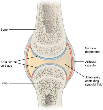
- Synovial joints consist of a joint cavity that possesses a thick fluid called synovial fluid.
- Synovial fluid cushions the ends of bones and reduces friction when we move our joints.
- A flexible articular capsule encloses and protects synovial joints.
Parts of synovial joints:
- A typical synovial joint consists of four essential structures, namely;
- Synovial cavity
- Articular cartilage
- Articular capsule
- Ligaments
1. Synovial cavity:
- Also known as joint cavity, synovial cavity is the space between two articulating bones.
- It contains folds of synovial membrane that sometimes contains pads of fat. These fatty pads help fill the spaces between articulating bones and also reduce friction.
- The synovial membrane secretes a thick synovial fluid that lubricates the synovial cavity.
- Synovial fluid gets its name because its thick consistency resembles the white of an egg.
- Combined with the articular cartilage, synovial fluid provides an almost friction-free surface for the easy movement of joints.
2. Articular cartilage:
- Articular cartilage is a hyaline cartilage which caps or covers the surface of bones facing the synovial cavity.
- It has a silvery-blue luster and appears as polished as a pearl.
- It acts like a shock absorber because of its thickness and elasticity.
- In certain joint diseases, articular cartilage gets worn away and restricts the movement of joints making it more painful.
- The cartilage itself is insensitive to feeling, since it has no nerve supply, but the other portions of the joint are supplied with pain-receptor nerve fibers.
- Several (not all) synovial joints have articular disks or fibro-cartilaginous disks.
- The role of such disks vary depending on the joints;
- They may act as shock absorbers to reduce the effect of shearing (twisting) on a joint.
- They prevent the jarring between the bones.
- They adjust the unequal articulating surfaces of the bones so that the surfaces fit together more evenly.
- In some joints, the fibro-cartilaginous disk forms a complete partition, dividing the joint into two cavities.
- In the knee joint, the fibro-cartilages, called the medial and lateral menisci, are crescent-shaped wedges that form incomplete partitions.
- These menisci serve to cushion as well as guide the articulating bones.
- Many athletes, especially sprinters and football players, tear these menisci which is commonly referred to as torn cartilages.
3. Articular capsule:
- Articular capsule lines the synovial cavity in the non-cartilaginous parts of the joint.
- This capsule if reinforced with collagenous fibers is called fibrous capsule. Fibrous capsule is lax and pliable, permitting considerable movement.
- The inner lining of the capsule is the synovial membrane, which extends from the margins of the articular cartilages.
- The outer layer of the capsule is a fibrous membrane which extends from bone to bone across a joint and reinforces the capsule.
- So called double-jointed people have loose articular capsules.
4. Ligaments:
- The portion of fibrous capsule reinforced by a thick layer of collagenous fiber is called ligament.
- Ligament joins or connects two articulating bones in a joint.
- They vary in shape and even in strength, depending on their specific roles.
- Most ligaments are considered inelastic, yet they are flexible enough to permit movement at the joints.
- Under excessive stress, ligaments will tear rather than stretch.
- Torn ligaments are extremely painful and are accompanied by immediate local swelling.
- In general, ligaments are strong enough to prevent any excessive movement and strain. A rich supply of sensory nerves helps prevent a person from over-stretching ligaments.
Two other structures associated with joints, but not parts of them are;
- Bursae
- Tendon sheaths
1. Bursae:
- Bursae (sing. bursa) are flattened sac like structures filled with synovial fluid.
- Bursae are found wherever it is necessary to eliminate the friction that occurs when a muscle or tendon rubs against another muscle, tendon or bone.
- They also cushion certain muscles and facilitate the movement of muscles over bony surfaces.
- The inflammation of bursae is called bursitis.
2. Tendon sheaths:
- Tendon sheath is a modification of bursa.
- It is a long cylindrical sac like structure filled with synovial fluid.
- It surrounds long tendons that are subjected to constant friction such as tendons of the wrist, palm and finger muscles.
- Like bursae, tendon sheaths reduce friction and permit tendons to slide easily.