
- A joint, also known as articulation is the place where two adjacent bones or cartilages meet.
- Although most joints are movable, some are not.
- Movable joints provide the mechanism that allows the body to move through coordination of nervous, skeletal and muscular systems.
- Joints are classified on two bases:
I. On the basis of their function (Degree of movement):
- This classification is based on the degree of movement of bones in a joint.
- According to this system, a joint may be;
- Immovable (Synarthrosis):
- Such joints don’t allow movement because bones are rigidly joined together.
- e.g. Manubriosternal joint, the joints between the skull bones (Sutures)
- Slightly movable (Amphiarthrosis):
- They allow limited or slight movement.
- e.g. Pubic symphysis of the pelvis, intervertebral joints
- Freely movable (Diarthrosis):
- They permit a great deal of movement.
- e.g. Elbow, shoulder and ankle joints
- Immovable (Synarthrosis):
II. On the basis of their structure:
- This classification is based on the presence or absence of a joint cavity and the kind of supporting tissue that binds the bones together.
- According to this system, there are three types of joints;
- Fibrous joints
- Cartilaginous joints
- Synovial joints
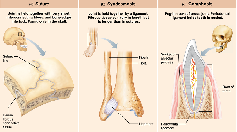
1. Fibrous joints:
- Lack a joint cavity
- Fibrous connective tissue unites the articulating bones tightly
- Mostly immovable and some are slightly movable
- Usually synarthroses
- They are of three types;
https://www.pinterest.com/
a. Sutures:
- Found only in the skull
- Fibrous tissue connects the articulating bones in children
- Bones are permanently fused in adults
- Some movement in fetuses and young children but immovable in adults
- e. g. Cranial sutures
b. Syndesmoses:
- Articulating bones are held together (without touching each other) by fibrous or interossoeus ligaments.
- Allow slight movement: twisting of forearm (pronation and supination)
- e.g. Inferior tibiofibular joint, interosseous ligament between shafts of radius and ulna
c. Gomphoses:
- A peg fits into a socket.
- Mostly immovable and some may have very slight movement of teeth in their sockets.
- e. g. roots of teeth in alveolar processes of mandible and maxillae
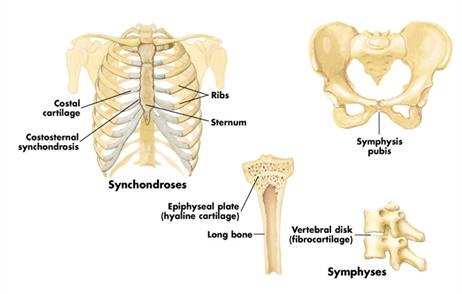
2. Cartilaginous joints:
- The articulating bones are united by a plate of hyaline cartilage or fibro-cartilaginous disk.
- They also lack a joint cavity.
- Most of them are slightly movable while some are immovable.
- Usually amphiarthroses
- They are of two types;
https://moberlyanatomy.weebly.com/
a. Synchondroses:
- Also called a primary cartilaginous joint.
- It is a temporary joint composed of an epiphyseal plate of hyaline cartilage that joins the diaphysis and epiphysis of a growing long bone.
- Chief function is to permit growth of the bones, not their movement.
- Immovable joints
- e. g. epiphyseal plate of femur, union of manubrium and the body of sternum
b. Symphyses:
- Also called secondary synchondrosis
- The two articulating bony surfaces are covered by thin layers of hyaline cartilage.
- Between them are disks of fibro-cartilage (collagenous fibers with cartilage cells) that serve as shock absorbers.
- Allow slight movement
- e. g. pubic symphysis, manubriosternal joint, intervertebral joints
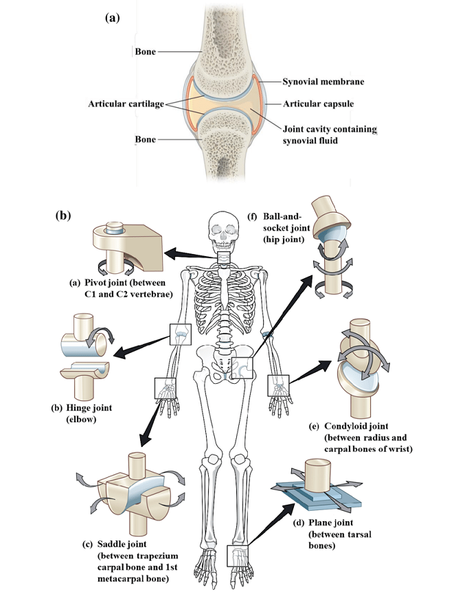
3. Synovial joints:
- The ends of the articulating bones are covered with a smooth hyaline articular cartilage and the joint is enclosed by a flexible articular capsule.
- Joint cavity is present which is also called a synovial cavity.
- The joint is lubricated by a thick fluid called synovial fluid.
- Articular capsules that are reinforced with collagenous fibers are called fibrous capsules, which are further reinforced at certain portions with collagenous fibers to form a ligament.
- Allow more free movements than any other type of joint, i.e. usually diarthroses
- Synovial joints are classified according to the shape of their articulating surfaces and the type of joint movements those shapes permit.
- Six types of synovial joints are recognized;
Also see in detail: Structure of a Typical Synovial Joint
https://www.researchgate.net/figure/1-a-A-Synovial-Joint-b-Types-of-Synovial-Joints-Image-source_fig1_341110002
a. Hinge joints:
- The convex surface of one bone fits into the concave surface of another bone.
- Allows uniaxial movement like flexion and extension (back and forth movement occurs around a single transverse axis)
- e. g. joints in elbow, interphalangeal joints in fingers, knee and ankle
b. Pivot joints:
- Central bony pivot is surrounded by a collar of bone and ligament.
- Allows uniaxial movement like supination, pronation and rotation around a central axis through the center of the pivot.
- e. g. proximal radioulnar joint, atlantoaxial joint
c. Ball and socket joints:
- Globe-like head of one bone fits into a cup-like concavity of another bone.
- This is the most freely movable of all the joints allowing multiaxial movement
- Allows movements like flexion, extension, medial (internal) rotation, lateral (external) rotation, abduction, adduction and circumduction.
- e. g. shoulder joint and hip joint
d. Condyloid or Ellipsoidal joints:
- They are the modifications of multiaxixal ball and socket joints.
- However, because the ligaments and muscles around the joint limit the rotation to two axes, the joint is classified as biaxial joint.
- Allows movements like flexion and extension of the hinge joints as well as abduction, adduction and circumduction.
- No rotational movement in permitted.
- e. g. metacarpophalangeal (knuckle) joints except thumb
e. Gliding joints (Plane joints):
- They are almost always small and are formed by essentially flat articular surfaces.
- One bone slides on another bone with a minimal axis of rotation, if any.
- Allows multiaxial movement like simple gliding within narrow limits.
- e. g. between articular processes of vertebrae, acromioclavicular joint, some carpal and tarsal bones
f. Saddle joints:
- The opposing articular surfaces of both bones are shaped like a saddle, i.e. they have both concave and convex areas that fit into one another at right angles to each other.
- Allows multiaxial movements like abduction, adduction, opposition and reposition.
- e. g. carpometacarpal joint of thumb