- The fusion of male gamete and female gamete in sexual reproduction is called fertilization.
- In angiosperms, the female gamete lies deep in the ovarian cavity, whereas the pollen grains after pollination are deposited on the stigma away from the egg.
- Therefore, for fertilization to occur, pollen grains have to travel a distance between the stigma and the ovarian cavity in order to reach to the egg.
You may also want to see: Sequential events and stages in the development of frog (pre-embryonic, embryonic and post-embryonic development)
Formation of pollen tube:
- Pollen grains deposited on stigma absorb stigmatic fluid and swell up.
- Sugar solution (stigmatic fluid) stimulates the germination of pollen grain. The exine ruptures and intine comes out as a pollen tube.
- Usually one pollen tube develops from a pollen grain. The pollen tube grows down through the stigma and style to the ovary.
- The pollen tube secretes some enzyme which dissolves the tissues of the stigma and the style and help it bore its way through to the ovary.
- Each pollen grain has two nuclei; a vegetative nucleus and a generative nucleus.
- The vegetative nucleus or tube nucleus is located at the growing tip and regulates the growth of the pollen tube.
- The generative nucleus divides by mitosis during migration towards the ovary and forms two male gametes.
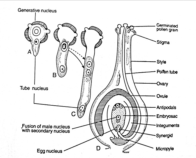
Entry of pollen tube into the ovule:
- After reaching the ovary, the pollen tube enters into the ovule through a micropyle and rarely through other regions. Based on the modes of pollen tubes entering into the ovule, fertilization is classified into 3 types:
- Porogamy: The pollen tube enters through the micropyle. E.g. Lily
- Chalazogamy: The pollen tube enters through the chalazal end. E.g. Casuarina
- Misogamy: The pollen tube enters through the integuments. E.g. Cucurbita, Populus
- The pollen tube penetrates the embryo sac by growing into one of the synergids which play an important role in directing the pollen tube growth by secreting some chemotropically active substances.
Double fertilization and triple fusion:
- The pollen tube bursts to discharge two male gametes inside the embryo sac. The tube nucleus is generally disorganized by this time.
- One of the two male gametes fuses with the egg cell (ovum) to form a diploid zygote.
- The other male gamete fuses with the polar nuclei to form a triploid nucleus or primary endosperm nucleus.
- The zygote on its further development forms the embryo, while the primary endosperm nucleus forms the endosperm.
- Fertilization of one male gamete with the egg and another male gamete with the polar nucleus is called double fertilization which is a special feature of angiosperms.
- It was discovered by Nawaschin in 1898.
- The fusion of the male gamete with the polar nuclei is called triple fusion as usually three nuclei take part in it.
Development of embryo:
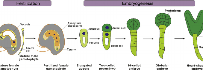
- A diploid zygote is the first cell of the embryo.
- In the earlier stages of embryo development, there are no fundamental differences between a dicot and a monocot embryo.
- The process of embryo development is similar up to the octant stage and differences appear with the plumule and cotyledon initials.
- Development of dicot embryo:
- The zygote divides by a transverse division to form two unequal cells;
- A large suspensor cell towards the micropylar end and
- A small embryonal cell towards the chalazal end.
- From this two-celled stage until the initiation of organs development, the embryo is called pro-embryo.
- The suspensor cell divides transversely to form a filament of 6-10 cells, called the suspensor.
- The end cell of suspensor which is called basal cell becomes bulbous and acts as absorbing organ for the growing embryo.
- The zygote divides by a transverse division to form two unequal cells;
-
- The suspensor elongates and pushes the developing embryo deeper into the endosperm to get nutrition.
- The cell of the suspensor lying next to the embryo is called hypophysis which forms roots later on.
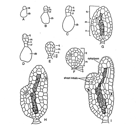
- The embryonal cell enlarges in size and becomes somewhat spherical.
- It divides by two longitudinal divisions at right angles to each other, resulting in the formation of 4-celled embryo, called quadrant stage.
- Each cell of the quadrant divides transversely to from eight celled structure or octant stage. The terminal or outer four cells of the octant are called epibasal cells while the four cells towards the suspensor are called hypobasal cells.
- The epibasal cells form the plumules and the cotyledons whereas the hypobasal cells from the hypocotyl.
- The embryonal cells divide further and gradually become heart shaped. The two lateral lobes continue to grow and form cotyledons. A small group of cells between these lobes form the plumule. The hypocotyl and the cotyledons soon elongate in size.
- Development of monocot embryo:
- The zygote divides transversely to form a basal cell and a terminal cell.
- The basal cell present towards the micropylar end doesn’t divide anymore but gradually becomes larger to form the most important part of the suspensor.
- The terminal cell gives rise to the whole of the embryo and a part of the suspensor. It divides to form a three-celled pro-embryo.
- The lower most terminal cell undergoes further longitudinal and transverse divisions giving rise to eight cells arranged in two tiers.
- The terminal cells by further growth and development gives rise to single terminal cotyledon.
- The middle cell of pro-embryo forms the hypocotyl, radicle and the rest of the suspensor.
- The plumule arises as a depression, midway between the bases of the cotyledon and the axis of the embryo and thus, it is lateral in position.
Structure of embryo:
- A typical dicot embryo comprises of an embryonal axis with two cotyledons.
- The portion of the embryonal axis above the level of cotyledons is called epicotyl and that below the level of cotyledons is known as hypocotyl.
- The epicotyl terminates into plumule and the hypocotyl bears the radicle at its lower end.
- The embryo of a monocot differs from that of a dicot mainly in having only one cotyledon.
Seed formation in monocot and dicot plants:
- An embryo with cotyledons, endosperm and seed coat is called a seed. The ovule forms the seed.
- As the embryo is formed, the integuments become lignified and form the seed coat. The outer integument forms the testa and the inner forms tegmen.
- The seed coat provides protection to the internal parts of the seed.
- In the development of embryo, the endosperm is used in some cases and food is stored only in cotyledons. Such seeds are called non-endospermic seeds. g. pea, bean, gram, Vallisnaria spp. etc. However, in other cases, the endosperm may be present after the development of the embryo, such seeds are called endospermic seeds. E.g. barley, cotton, coffee, maize etc.