
- The process of respiration in human beings gets completed in four steps;
- Breathing
- External respiration
- Internal respiration (tissue respiration)
- Cellular respiration (Oxidation of glucose)
- Breathing is a physical process of taking in oxygen from the atmosphere and throwing out carbon-dioxide with the help of our respiratory tract. i.e. The process of inhalation and exhalation of O2 and CO2 respectively is known as breathing.
- Lungs are the principal organs of our respiratory system.
- They consist of millions of minute air sacs called alveoli which help in the exchange of gases between the blood capillaries and lungs.
- Different muscles present in and around the thoracic and abdominal cavity help in the process of breathing. Such muscles are: Diaphragm, External intercostal muscles, Internal intercostal muscles and abdominal muscles.
- Breathing further involves two steps:
- Inhalation or inspiration:
- It is an active process by which fresh air is taken in from the atmosphere.
- Diaphragm becomes flat and gets lowered by the contraction of its muscle fibers thus increasing the volume of thoracic cavity in length.
- External intercostal muscles: They occur between ribs. These muscles contract and pull the ribs and sternum upward and outward thus increasing the volume of the thoracic cavity.
- Abdominal Muscles: These muscles relax and allow the compression of abdominal organs by the diaphragm.
- There will be an increase in intrapulmonary volume and decrease in intrapulmonary pressure (759 mmHg, difference of 1 mmHg), in relation to atmospheric pressure (760 mmHg taken as standard atmospheric pressure).
- Oxygen flows into the lungs (from an area of high pressure gradient to low pressure) until intrapulmonary pressure becomes equal to the atmospheric pressure.
- Exhalation or expiration:
- It is a passive process by which the foul air is expelled out from the lungs.
- The muscle fibers of diaphragm relax making it convex, decreasing volume of thoracic cavity.
- External intercostal muscles relax and bring the ribs and sternum to the original position. This decreases the volume of thoracic cavity.
- Contraction of the abdominal muscle compresses the abdomen and pushes its content towards the diaphragm.
- Intrapulmonary volume decreases and intrapulmonary pressure increases (difference of 1 mmHg).
- So, gases move out into the atmosphere, down the pressure gradient until intrapulmonary pressure becomes equal to the atmospheric pressure.
- This cycle continues with inhalation and exhalation alternating with each other.
- Inhalation or inspiration:

Neural Regulation of Breathing:
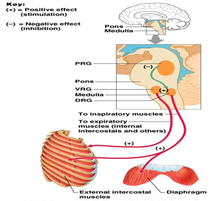
- The process of breathing is controlled by the respiratory centers present in brain which is located in the medulla oblongata and pons, in the brainstem.
- The respiratory centers is made up of three major respiratory groups of neurons, two in medulla and one in pons.
- In medulla, they are the dorsal and the ventral respiratory groups whereas in the pons, the pontine respiratory group includes two areas known as the pneumotaxic center and the apneustic center.
Control centers in the Medulla (Medullary regulatory center):
- It is called a rhythmicity center also known as medullary rhythmicity center where the rhythmic cycle of breathing originates.
- It rhythmically stimulates inspiratory muscles to contract which will result in resting respiration.
- It consists of further two groups;
1. Dorsal respiratory groups (DRG): It consists of inspiratory neurons that control the basic rhythm of breathing by triggering inspiratory impulses.
- These neurons send impulses to the motor nerves of diaphragm and external intercostal muscles.
- DRG nerves extend into the Ventral respiratory group (VRG), but the VRG neurons do not extend into the DRG.
- The sensory impulses from the lungs, airways, peripheral chemoreceptors, and joint proprioceptors (a sensory receptor which receives stimuli from within the body, especially one that responds to position and movement) are brought to the DRG by Vagus and glossopharyngeal nerves.
- They send neural stimuli via nerves to inspiratory muscles, to contract for inspiration to occur i.e send impulses to the external intercostal muscles and the diaphragm at regular rhythm, so that we are breathing at the rate 12-15 breaths/ min. When it shuts off, expiration occurs passively.
2. Ventral respiratory group (VRG): It contains both inspiratory and expiratory neurons.
- It works together with and in the same manner as DRG to initiate normal resting inspiration.
- Expiratory neurons stimulate the internal intercostal muscles required during forced expiration.
- VRG sends inspiratory impulses to the muscles of larynx, pharynx, diaphragm and external intercostal muscles.
- Other VRG neurons send expiratory signals to abdominal muscles and internal intercostal muscles.
Control centers in the Pons:
- Pons moderates the rhythm (to increase or decrease rhythm) of breathing.
- It helps in easy smooth transition between inspiration & expiration (between phases of breathing). It has 2 centers;
1. Apneustic center:
- It helps in process of inspiration.
- It helps stimulate medulla (both DRG and VRG) to keep stimulating inspiratory muscles.
- Over stimulation from the apneustic center results in apneustic breathing which is characterized by long gasping inspirations interrupted by occasional expirations.
2. Pneumotaxic center:
- It inhibits inspiration.
- It sends inhibitory signals to the inspiratory centers of the medulla and limits the inspiratory volume and rate (duration), so that the lungs are not over exerted (shortness of breathing).
Respiratory rate:
- A person’s respiratory rate is the number of breaths taken per minute which is also known as a breathing rate.
- The normal respiration rate for an adult at rest is 12 to 20 breaths per minute.
- A respiration rate under 12 or over 25 breaths per minute while resting is considered abnormal.
Regulation of respiratory rate:
1. By Peripheral Chemoreceptors:
- They are located in the carotid and aortic bodies and they notice any chemical changes in blood, oxygen, carbon dioxide, pH, and react accordingly.
- They send the impulses to the medulla to increase the RR and correct the abnormalities and also maintain normal homeostasis.
2. By Central chemoreceptors:
- They are present in medulla are highly sensitive to high level of carbon dioxide.
- Carbon dioxide crosses the blood brain barrier (BBB) which results in acidosis and finally detected by chemoreceptors.
- This will lead to increased RR to correct the high level of Carbon dioxide and acidosis i.e. increased ventilation.
3. Higher centers regulating Respiratory rate (RR):
- Cerebral cortex: It has a role in voluntary breathing.
- Limbic system (emotional brain): During excitement and fear, we breathe faster. This is because limbic system stimulates respiratory centers to work at higher rate.
- Hypothalamus: During fever RR increases.

Mechanism of breathing and its neural regulation