
Chromosomes
- The nucleus of eukaryotic cells consists of a thin reticulate complex of DNA and proteins (histone), which are not distinctly visible in a non-dividing cell. These thread like structures are called chromatin fibers or chromatin reticulum.
- When a cell starts dividing, the chromatin fibers become shorter, thicker and more distinct inside the cell. These microscopic short, rod like structures that carry hereditary information in the form of genes are called chromosomes.
- After the metaphase stage of cell division, each chromosome divides or splits longitudinally into two thick and short strands which are distinctly visible inside the cell. Each of these two strands is called a chromatid.
Structure of chromosome:
- The chromosomes vary widely between different organisms. Eukaryotic cells have large number of linear chromosomes (rod-like) inside the nucleus and cells of prokaryotes have smaller and circular DNA in their cytoplasm.
- Each eukaryotic cell consists of a long DNA molecule surrounded by histone proteins in the condensed form inside the nucleus.
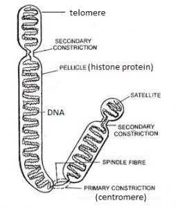
- Each chromosome has a constricted structure in it called the primary constriction or centromere, which holds together the two sister-chromatids (the daughter strands of a replicated chromosome).
- A telomere is a region at each end of a chromosome, which protects the end of the chromosome from deterioration or from fusion with neighboring chromosomes.
- Besides the primary constriction, in certain chromosomes there is/are secondary constriction(s) as well. Some chromosome consists of a small pinched off portion from the chromosomal body called a ‘satellite’ and the chromosome is called a SAT chromosome.
Types of chromosome:
1.On the basis of position of centromere:
a.Metacentric chromosome: The chromosome in which the centromere is exactly at the center. Human chromosomes 1 and 3 are metacentric.
b.Sub-metacentric chromosome: The chromosome in which the centromere is located towards the center but not exactly at the center. Human chromosomes from 4 through 12 are sub-metacentric.
c. Acrocentric chromosome: The centromere lies towards the pole or away from the center of the chromosome leading to one very long and one very short section or arm. Human chromosomes 13, 15, 21 and 22 are acrocentric chromosomes.
d.Telocentric chromosome: The centromere lies at the very end or pole of the chromosome. Humans don’t possess telocentric chromosomes but are found in some other species like mice.
2.On the basis of function:
a. Autosomes: Those chromosomes which transfer somatic hereditary characteristics from parents to the offspring and are not responsible for the determination of gender (sex) are called autosomes. In human beings, 22 pairs (from 1 to 22) of autosomes are present.
b.Sex chromosomes: Sex chromosomes are those chromosomes that are needed for the determination of sex (male or female) of an individual. In human beings, the last one pair of chromosomes (XY) are sex chromosomes.
Types of cells on the basis of number of chromosomes:
- Haploid cells: The cells with only set of chromosomes (either from father or mother) are called haploid cells. There is no homologous pairing of chromosomes in a haploid cell. E.g. gametes. They are represented by ‘n’ or ‘x’.
- Diploid cells: The cells with two sets of chromosomes (one from father and the other from mother) in the form of homologous pair are called diploid cells. E.g. Zygote formed by the fusion of gametes, somatic cells. They are represented by ‘2n’ or ‘2x’.
Homologous chromosomes:
Homologous chromosomes are the pairs of chromosomes (one from each parent) that are similar in length, gene position, and centromere location. The position of the genes on each homologous chromosome is the same, however the genes may contain different alleles. Homologous chromosomes are always present in the diploid cells.
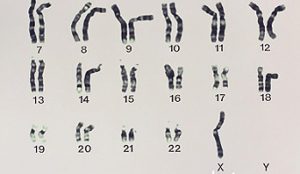
Karyotype:
The complete set of chromosomes in a species or in an individual organism which are arranged on the basis of their length, the position of the centromeres, banding pattern, any differences between the sex chromosomes, and any other physical characteristics is called a karyotype. In general karyotype is the number and appearance of chromosomes inside the nucleus of eukaryotic cells.
Chromosomal disorders:
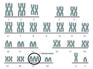
Aneuploidy (an abnormal number of chromosomes) occurs when an individual either is missing a chromosome from a pair (monosomy) or has more than two chromosomes of a pair (trisomy, tetrasomy, etc.) and this results to chromosomal disorders, symptoms of which can be distinctly observed.
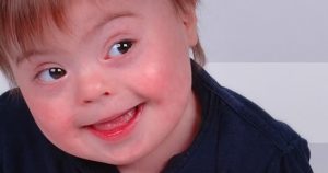
1.Down syndrome:
- is one of the most common autosomal abnormalities in humans, occurs in 1/1000 babies born each year
- It is named after John Langdon Down, the British doctor who fully described the syndrome in 1866.
- Occurs because of an extra chromosome (either all or part of the third chromosome) in the 21st pair (trisomy) in which an abnormal egg has been fertilized by a normal sperm.
- The total number of chromosomes in such individual is 47.
- Symptoms include:- round face, mental impairment, stunted growth , slanted eyes, short neck, protruding tongue, outer ears, congenital heart disease, flattened nose, separation of first and second toes etc.
- Since the patient looks like a Mongolian, also called a mongoloid disease
- There is no cure for Down syndrome but we can improve their quality of life by education and proper care.
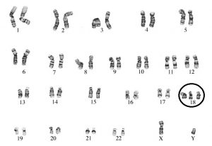
2. Edwards syndrome:
- It is an autosomal disorder and occurs because of an extra chromosome (either all or part of the third chromosome) in the 18th pair (trisomy).
- It occurs in around one in 5,000 live births
- It is named after John Hilton Edwards, who first described the syndrome in 1960 and many of those affected die before birth.
- The total number of chromosomes in such individual is 47.
- Symptoms include cleft lip/cleft palate, upturned nose, narrow eyelid folds, widely spaced eyes, small head, intestines protruding outside the body etc.
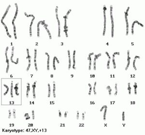
3. Patau syndrome:
- It is an autosomal disorder in which some or all of the cells of the body contain extra chromosome in the 13th
- The extra genetic material disrupts normal development, causing multiple and complex organ defects.
- The chromosomal nature of the disease was ascertained by Dr. Klaus Patau in 1960 and hence named on his honor.
- Patau syndrome affects somewhere between 1 in 10,000 and 1 in 21,700 live births.
- 47 chromosomes are present in such individuals.
- Symptoms include:- prominent heel, deformed feet, intellectual disability and motor disorder, small head, structural eye defects etc.
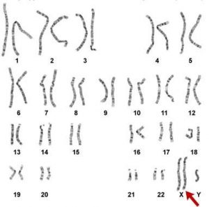
4. Klinefelter syndrome:
- is the set of symptoms that result from two or more X chromosomes in males. i.e it is a disorder in the sex chromosomes and represented by XXY.
- Occurs when a normal egg (X) is fertilized by an abnormal sperm (XY), or a normal sperm (Y) fuses with an abnormal egg (XX).
- It is named after Harry Klinefelter who identified the condition in the 1940s and occurs in 1:500 to 1:1,000 live male births.
- In addition to a Y chromosome there is an extra X chromosome such that there is a total of 47 chromosomes rather than the usual 46 in such males.
- Symptoms include:- sterility and small testicles, less body hair, breast growth, less interest in sex etc.
5. Turner syndrome:
- is a condition in which a female is partly or completely missing an X chromosome hence represented as XO or 45.
- While most people have 46 chromosomes, people with this syndrome usually have 45chromosomes.
- Henry Turner first described the condition in 1938 and later known to be the disorder in the sex chromosomes.
- Turner syndrome occurs in between one in 2000 to one in 5000 females at birth
- Generally people with Turner syndrome have a shorter life expectancy, mostly due to heart problems and diabetes.
- Symptoms include:- Short stature, broad chest and widely spaced nipples, low posterior hairline, low-set ears, reproductive sterility, underdeveloped ovaries, the absence of a menstrual period etc.
There shall have been notes on honeybee and silkworm as well..
thank you..will post that as soon as possible 🙂