- The human skeleton has two major divisions; the axial skeleton and the appendicular skeleton.
- The axial skeleton forms the median longitudinal axis of the body that consists of the skull, vertebral column, sternum and the rib cage.
- The appendicular skeleton is composed of the upper and the lower extremities which include the pectoral and the pelvic girdles.
- These girdles (pectoral and pelvic) attach the upper and lower appendages or limbs to the axial skeleton.
The axial skeleton:
- The axial skeleton consists of the skull, vertebral column and the rib cage (with sternum).
- At the top of the axial skeleton is the skull (the skeleton of the head).
The skull (29 bones):
- It protects the brain forming a hard box of bones outside the brain.
- The skull consists of 29 bones (including the ear ossicles, mandible and hyoid).
- The skull can be divided into:
- The cranial bones (cranium)
- The facial bones
- The cranim consists of the following 8 bones:
- Frontal (1 bone): Anterior and superior part of cranium, forehead and brow areas, which protects front part of the brain.
- Parietal (2 bones): Superior sides and roof cranium, between frontal and occipital bones, that protects the top and sides of the brain.
- Temporal (2 bones): sides and base of cranium at temples that protect the ear ossicles.
- Occipital (1 bone): Posterior part of the cranium including the base that protects the posterior part of the brain.
- Ethmoid (1 bone): Base of cranium, anterior to the body of sphenoid and forms the roof of nasal cavity and septum.
- Sphenoid (1 bone): Base of cranium, anterior to occipital and temporal bones which houses the pituitary gland.

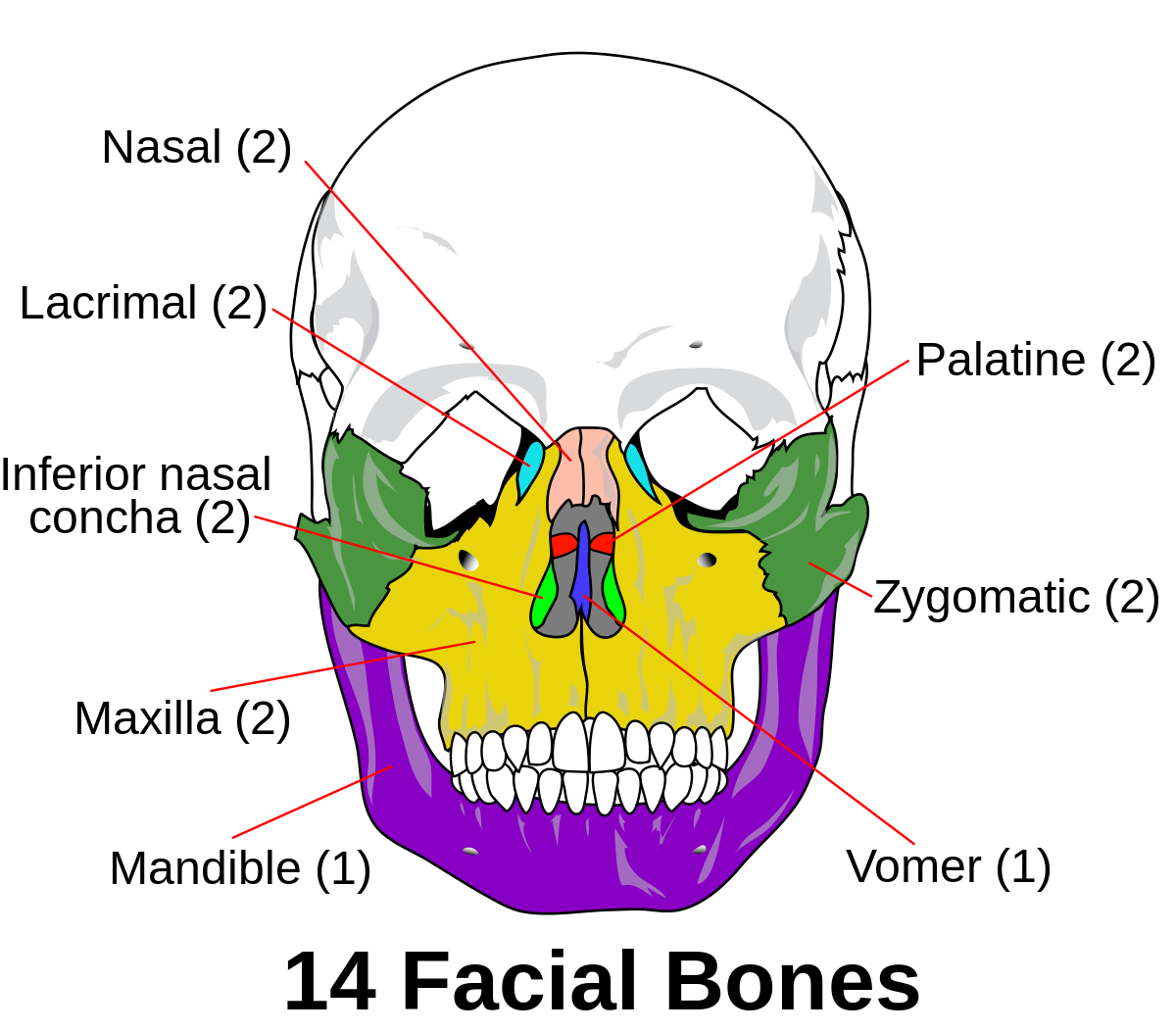
- The facial bones consists of the following 14 bones present in the face:
- Lacrimal (2 bones): Small, thin and rectangular, form the medial wall of eye orbits.
- Nasal (2 bones): Small, oblong and form the upper bridge of nose, attached to a nasal cartilage.
- Inferior nasal conchae (2 bones): Thin, shaped like curved leaves, form the lateral walls of nasal cavities.
- Zygomatic (2 bones): Cheek bones below and lateral to orbits.
- Vomer (1 bone): Thin, forms the posterior and inferior part of nasal septum.
- Maxillae (2 bones): Upper jaw and anterior part of the hard palate.
- Palatine (2 bones): L-shaped, posterior part of hard palate and the floor of nasal cavity and orbit.
- Mandible (1 bone): Largest and strongest facial bone, lower jaw, extending from chin to mandibular fossa of temporal bone.

- Other skull bones:
- Hyoid (1 bone): U-shaped, below the root of tongue and above the larynx, site of attachment for some muscles that help in speaking and swallowing.
- Ossicles of ear (6 bones): Incus (2), Malleus (2) and Stapes (2) Lying inside the cavity of temporal bone that convey sound vibrations from eardrum to the inner ear.
The vertebral column (26 bones):
- It is commonly called the spine or the spinal column which is a roughly S-shaped column of bones.
- It extends from the base of the skull through the entire length of the trunk.
- This column consists of 26 separate bones called vertebrae.
- The vertebral column is stabilized by ligaments, muscles and intervertebral disks to permit twisting and bending movements.
- The vertebrae in the vertebral column are arranged as follows:
- Cervical vertebrae (7 bones): First (Atlas), second (Axis) and seventh vertebrae are modified; third through sixth are typical, support head and allow movement of neck.
- Thoracic vertebrae (12 bones): Bodies and transverse processes have facets that articulate with the ribs; allow some movement of spine in the thoracic region.
- Lumbar vertebrae (5 bones): Largest, strongest vertebrae, allow forward and backward bending of spine
- Sacral vertebrae (1 bone): Wedge-shaped, made up of 5 fused bones, support the vertebral column and give strength and stability to the pelvis.
- Coccygeal vertebrae (1 bone): Triangular tail bone (4 bones fused to form 1), united with the sacrum by intervertebral disk.

* The vertebral column of infants consists of 33 bones, in which sacral region has 5 bones and coccygeal region has 4 bones which are not yet fused.
The sternum and the rib cage (25 bones):
- The cage-like thoracic skeleton protects the heart, the lungs and some abdominal organs.
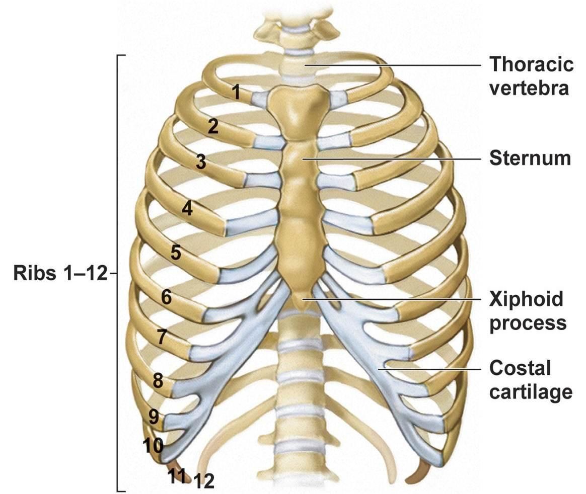
- The rib cage anteriorly consists of 12 pairs of ribs, 12 costal cartilages and the sternum.
- The sternum or the breast bone (1 bone): The midline bony structure of the anterior chest wall.
- It resembles a dagger in the adult that consists of a manubrium, a body and xiphoid process.
- Ribs are long, curved bones, varying in length and width. With costal cartilages,they provide flexible mechanism for breathing and support bones of upper extremities.
- 12 pairs of ribs altogether can be divided into 3 types:
- True ribs (1st-7th pairs): They arise from the thoracic vertebrae at the back and attach directly to the sternum in the front.
- False ribs (8th -10th pairs): They arise from the thoracic vertebrae at the back but don’t attach to the sternum directly, instead attach to the cartilage of the 7th true rib.
- Floating ribs (11th and 12th pairs): They are attached only to the vertebral column at the back and nothing in the front.
