
- The human eye is the organ of vision and has the ability to form images of the outside world.
- They are located and protected in deep sockets of the skull, called orbits.
- Each eyeball is held in position and moved within the orbit by six distinct set of muscles attached to its outer surface.
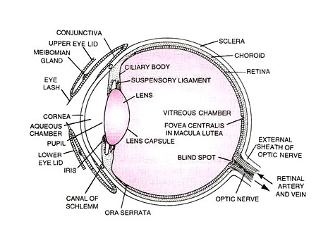
- The eyeball is a sphere of about 2.5 cm diameter and consists of three concentric layers, namely;
- The outer supporting layer
- The middle vascular layer
- The inner retinal layer
1. Supporting layer of eyeball:
- It consists of two parts; sclera and cornea
Sclera:
- It is the white part of the eye which is an opaque layer extending posteriorly over five-sixths of the outer layer of the eyeball.
- It is made up of very strong and non-elastic fibrous connective tissues.
- It gives eyeball its shape and protects the inner layers of eyeball.
Cornea:
- Sclera slightly bulges anteriorly (front part of the eye) which is called cornea.
- It is transparent and non-vascular.
- It acts as a non-adjustable lens through which light enters into the eyeball.
- A thin transparent membrane behind the eyelids called conjunctiva terminates into the cornea.
2. Vascular layer of eyeball:
- The second or middle layer is the choroid layer which is thin and vascular (has blood vessels to supply blood and oxygen) made up of connective tissue.
- It lies in between sclera and retina.
- This layer consists of various parts as follows:
Ciliary body:
- It is a thickened vascular layer in the anterior portion of the eyeball.
- These muscles help lens to focus by either increasing or decreasing tension on suspensory ligament of lens.
- It also produces aqueous humor and some elements of vitreous humor.
Ciliary processes:
- These are inward extensions (muscles) of ciliary body that hold the lens in place.
Iris:
- This is the colored part of eye which is an anterior extension of the choroid layer.
- It is thin and muscular which regulates the size of pupil, and thus amount of light entering the eye.
- The contraction and relaxation of iris is a reflex response.
Lens:
- It is elastic, colorless and transparent biconvex body made up of epithelial cells lying posterior to iris.
- The lens can accommodate and change its shape, focusing on different objects at different distances, i.e. the lens is adjustable.
- This accommodation is brought about by ciliary muscles.
3. Retinal layer of eyeball:
- It is a multilayered, light-sensitive membrane which is the innermost layer of the eyeball.
- It is connected to the brain by optic nerve and consists of thicker neural layer (neuroretina) and thinner pigmented layer.
- This pigmented layer prevents the reflection of light from back of retina.
- Retina receives the focused light waves and transduces them into nerve impulses that the brain converts into visual perceptions.
- Each eye has about 125 million rods and 7 million cones in the retinal layer.
- Most of the cones are concentrated in the center of the retina directly behind the lens in an area called the macula lutea or yellow spot, especially in a small depressed rod-free area called the fovea or fovea centralis.
- The portion of the retina where the optic nerve exits from the eyeball and contains neither rods nor cones is called the blind spot or optic disk. This spot is not sensitive to light.
Two types of photoreceptor cells present in retina are:
Rod cells:
- Rods are sensory cells for perception of black to white shades.
- It functions in dim light and helps in night vision.
- It contains a photosensitive pigment called rhodopsin synthesized from vitamin A.
Cone cells:
- Cones are sensory cells for perception of colors.
- It functions in bright light and differentiates colors.
- It contains a photosensitive pigment called iodopsin.
Chambers of the eyeball:
- The eyeball is divided into 2 chambers by the lens.
Aqueous chamber:
- The region between the cornea and the lens is the aqueous chamber.
- Aqueous chamber is further divided into anterior chamber (between cornea and iris) and posterior chamber (between iris and lens).
- It is filled with a thin, watery fluid called aqueous humour (ultrafiltrate of blood) containing amino acids, glucose, ascorbic acid, hyaluronic acid and respiratory gases.
- The aqueous humour nourishes the lens and cornea, refracts light rays to focus on retina, and also maintains a constant pressure within the eyeball.
Vitreous chamber:
- It is the largest chamber in the eyeball (occupying 80% of the eyeball) present between the lens and the retina.
- It is filled with a more viscous, jelly-like or gelatinous vitreous humor, containing salts and muco-proteins.
- It keeps the eyeball from collapsing, supports retina and refracts light to focus on retina.