
The human heart
In all vertebrates including human beings, there is a single heart which acts as a pumping organ of blood vascular system. It receives and pumps blood from and to different organs of the body. For this purpose, it undergoes spontaneous, rhythmical and ceaseless contractions (beats) throughout the life.
- Appearance and position:
- Is a hollow, muscular organ (made up of involuntary cardiac muscles), roughly of the size of one’s fist (12×9 cm)
- Average weight is about 300gm in males and 250gm in females
- Is reddish-brown in color and somewhat conical in shape
- Located almost in the middle of the thoracic cavity close to its front wall and between the lungs. Its broad base faces upward and backward whereas the narrow apex is directed downward, forward and slightly to the left.
- Protective covering:
- The heart is enclosed in a tough, two-layered sac, the pericardium, comprising inner visceral pericardium attached to the heart and the outer parietal pericardium.
- The two layers have a potential space or cavity in between them, the pericardial cavity which consists of about 50 ml of pericardial fluid.
- This fluid keeps the heart moist, allows its free movement and reduces the friction between the heart wall and the surrounding tissues when the heart beats.
- The pericardium protects the heart from mechanical injury and checks its overstretching and overfilling with blood.
- External structure:
- The smaller upper chambers, auricles (atria) are demarcated externally from the lower larger chambers ventricles by an irregular groove called the coronary sulcus.
- The two ventricles are demarcated externally from each other by an oblique groove termed as inter-ventricular sulcus. This groove contains coronary blood vessels that supply blood to the heart muscles.
- Internal structure: The heart is a hollow organ that contains four chambers, various apertures, valves and great blood vessels.
a) Chambers of the heart:
- The upper smaller chambers of the heart are auricles or atria.
- They are the collecting chambers for the blood returning to the heart.
- They have thin walls, because they have to force blood into the ventricles that lie just below them.
- The two auricles are separated from each other by a partition, the inter-auricular septum or inter-atrial septum.
- The lower larger chambers of the heart are ventricles.
- They are the distributing chambers of the blood reaching from the auricles.
- The two ventricles are separated from each other by a partition, the inter-ventricular septum.
- The ventricles have thicker walls than that of the auricles
- The wall of the left ventricle is about three times as thick as that of the right ventricle, which is because the left ventricle has to pump the blood to the farthest end of the body whereas the right ventricle has to pump blood to the lungs, which lie nearby.
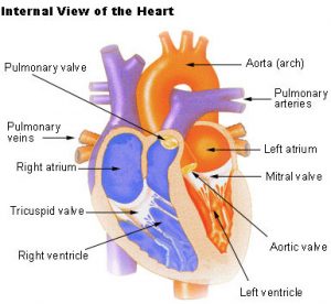
b) Great vessels, apertures and valves of the heart: The four heart valves allow the blood to flow through the heart in only one direction; they also prevent the back flow of blood.
i) Venae cavae:
- The right auricle receives two large veins: superior venacava and inferior vena cava.
- The superior venacava brings deoxygenated blood from the head and upper body parts.
- The inferior venacava returns the deoxygenated blood from the lower body parts.
ii) Right auriculo-ventricular aperture (A-V aperture) and tricuspid valve:
- The right auricle opens into the right ventricle through a wide passage, the right auriculo-ventricular aperture which is guarded by a one-way valve called the tricuspid valve.
- The valve consists of three membranous flaps which prevent the backflow of blood from right ventricle back to the right auricle during the contraction of the right ventricle.
iii) Pulmonary aorta and pulmonic semilunar valves:
- From its upper left corner, the right ventricle gives off a large blood vessel called the pulmonary arch or pulmonary artery.
- It divides into right and left pulmonary arteries that carry deoxygenated blood to the lungs for oxygenation.
- At the base of the pulmonary arch are three membranes, pocket-shaped flaps, the pulmonic semilunar valves.
- Pulmonic semilunar valves prevent the return of blood to the right ventricle.
iv) Pulmonary veins:
- The left auricle receives four pulmonary veins, two from each lungs.
- They bring oxygenated blood from the lungs.
- They don’t have any valves.
v) Left auriculo-ventricular aperture and bicuspid valve:
- The left auricle opens below into the left ventricle by a large passage, the left auriculo-ventricular aperture which is guarded by a one-way valve known as bicuspid valve or mitral valve.
- The valve consists of two membranous flaps which prevent the backflow of blood from left ventricle back to the left auricle during the contraction of the left ventricle.
vi) Systemic aorta and systemic semilunar valves:
- At its upper right angle, the left ventricle gives off a large blood vessel called the systemic aorta consisting of three regions; ascending aorta, arch of aorta and the descending aorta.
- At the base of the ascending aorta are three membranous, pocket-shaped systemic semilunar valves that check the return of the blood to the ventricle.
- Blood circulation (flow of blood through the heart):
- The sinu-auricular node (SA node) also called a natural pacemaker, initiates a wave of contraction which spreads over the walls of auricles and the ventricles.
- Because of the electrical impulse initiated by the SA node, the heart chambers undergo alternate contraction called systole and relaxation called diastole to pump the blood to and from different parts of the body.
- The superior and inferior venacavae carry deoxygenated blood from the upper and lower body parts respectively to the right auricle.
- From the right auricle, the blood passes into the right ventricle through the tricuspid valve which prevents the backflow of blood into the right auricle.
- The blood is pumped into the pulmonary trunk from the right ventricle which then divides into two pulmonary arteries (left and right pulmonary arteries) and carry blood to the lungs for oxygenation.
- The opening of the pulmonary aorta is guarded by three semilunar valves preventing the backflow of blood into the right ventricle.
- In the lungs, there is gaseous exchange and blood receives oxygen releasing carbon dioxide into the lungs.
- The oxygenated blood is carried from each lung to the left auricle by four pulmonary veins (two from each lung).
- The oxygenated blood from left auricle now passes to the left ventricle through bicuspid or mitral valve which prevents the backflow of blood into the left auricle.
- From the left auricle, the blood is now pumped into the systemic aorta guarded by three semilunar valves at the opening.
- Subsequently, the blood is now taken to different parts of the body by arteries.
Pulmonary circulation: The circulation of deoxygenated blood from right ventricle to the lungs by pulmonary arteries and oxygenated blood from lungs back to the heart by pulmonary veins is called pulmonary circulation of blood.
Systemic circulation: The circulation of oxygenated blood from left ventricle to different parts of the body (except the lungs) by systemic aorta with its branches and deoxygenated blood from different parts of the body to the right auricle of heart by superior and inferior venacavae is called systemic circulation of blood.
* Blood circulates twice through the heart before completing one full circuit, hence called double circulation.
